Featured Topics
Featured series.
A series of random questions answered by Harvard experts.

Explore the Gazette
Read the latest.

Kempner AI cluster named one of world’s fastest ‘green’ supercomputers

How humans evolved to be ‘energetically unique’

‘Harnessing evolution’
Epic science inside a cubic millimeter of brain.
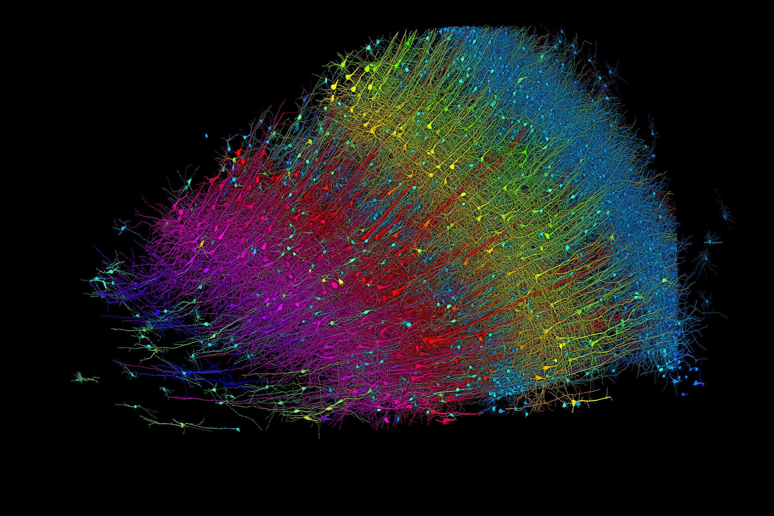
Six layers of excitatory neurons color-coded by depth.
Credit: Google Research and Lichtman Lab
Anne J. Manning
Harvard Staff Writer
Researchers publish largest-ever dataset of neural connections
A cubic millimeter of brain tissue may not sound like much. But considering that that tiny square contains 57,000 cells, 230 millimeters of blood vessels, and 150 million synapses, all amounting to 1,400 terabytes of data, Harvard and Google researchers have just accomplished something stupendous.
Led by Jeff Lichtman, the Jeremy R. Knowles Professor of Molecular and Cellular Biology and newly appointed dean of science , the Harvard team helped create the largest 3D brain reconstruction to date, showing in vivid detail each cell and its web of connections in a piece of temporal cortex about half the size of a rice grain.
Published in Science, the study is the latest development in a nearly 10-year collaboration with scientists at Google Research, combining Lichtman’s electron microscopy imaging with AI algorithms to color-code and reconstruct the extremely complex wiring of mammal brains. The paper’s three first co-authors are former Harvard postdoc Alexander Shapson-Coe, Michał Januszewski of Google Research, and Harvard postdoc Daniel Berger.
The ultimate goal, supported by the National Institutes of Health BRAIN Initiative , is to create a comprehensive, high-resolution map of a mouse’s neural wiring, which would entail about 1,000 times the amount of data the group just produced from the 1-cubic-millimeter fragment of human cortex.
“The word ‘fragment’ is ironic,” Lichtman said. “A terabyte is, for most people, gigantic, yet a fragment of a human brain — just a minuscule, teeny-weeny little bit of human brain — is still thousands of terabytes.”

Jeff Lichtman.
Kris Snibbe/Harvard Staff Photographer
The latest map contains never-before-seen details of brain structure, including a rare but powerful set of axons connected by up to 50 synapses. The team also noted oddities in the tissue, such as a small number of axons that formed extensive whorls. Because the sample was taken from a patient with epilepsy, the researchers don’t know whether such formations are pathological or simply rare.
Lichtman’s field is connectomics, which seeks to create comprehensive catalogs of brain structure, down to individual cells. Such completed maps would unlock insights into brain function and disease, about which scientists still know very little.
Google’s state-of-the-art AI algorithms allow for reconstruction and mapping of brain tissue in three dimensions. The team has also developed a suite of publicly available tools researchers can use to examine and annotate the connectome.
“Given the enormous investment put into this project, it was important to present the results in a way that anybody else can now go and benefit from them,” said Google collaborator Viren Jain.
Next the team will tackle the mouse hippocampal formation, which is important to neuroscience for its role in memory and neurological disease.
Share this article
You might like.
Computational power can be used to train and run artificial neural networks, creates key advances in understanding basis of intelligence in natural and artificial systems
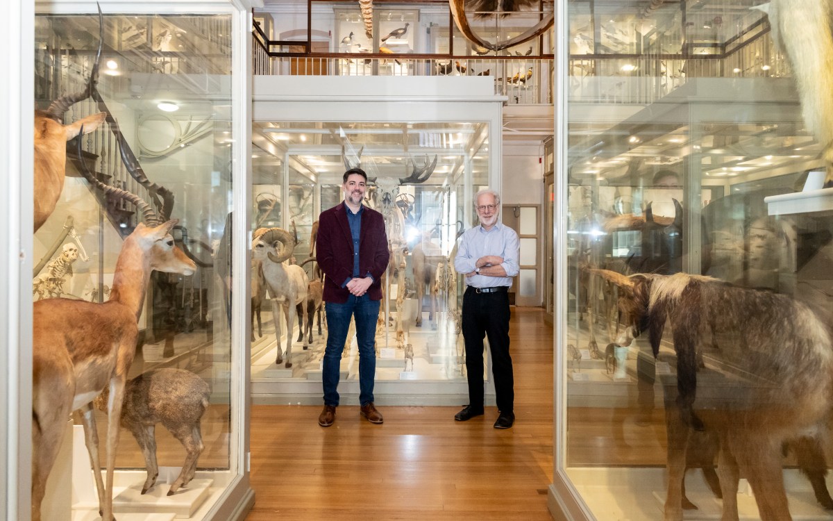
Metabolic rates outpaced ‘couch potato’ primates thanks to sweat, says new study
New tool allows researchers to study gene mutation directly within living human cells
8 Harvard students named Rhodes Scholars
5 in U.S. class, most for any institution, joined by 3 international recipients
Too much sitting hurts the heart
Even with exercise, sedentary behavior can increase risk of heart failure by up to 60%, according to study
Is cheese bad for you?
Nutritionist explains why you’re probably eating way too much
Thank you for visiting nature.com. You are using a browser version with limited support for CSS. To obtain the best experience, we recommend you use a more up to date browser (or turn off compatibility mode in Internet Explorer). In the meantime, to ensure continued support, we are displaying the site without styles and JavaScript.
- View all journals
Brain articles from across Nature Portfolio
The brain is the part of the central nervous system that is contained within the skull. It is responsible for executive and cognitive functions and regulates the functioning of the other parts of the nervous system.
Latest Research and Reviews
Microstructural asymmetry in the human cortex
The human cortex displays an anterior-to-posterior asymmetry, identified via both post-mortem and in vivo microstructural measurements. Microstructural asymmetry is heritable, varies across cortical layers and between sexes, and relates to functional asymmetry and behavior.
- Amin Saberi
- Sofie L. Valk
Extrauterine support of pre-term lambs achieves similar transcriptomic profiling to late pre-term lamb brains
- Jennifer L. Cohen
- Felix De Bie
- Alan W. Flake
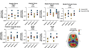
Restoring brain connectivity by phrenic nerve stimulation in sedated and mechanically ventilated patients
Bassi et al investigate the impact of phrenic nerve stimulation on deeply sedated, mechanically ventilated patients with acute respiratory distress syndrome. Cortical activity, connectivity, and synchronization are increased when phrenic stimulation is included in addition to invasive mechanical ventilation.
- Thiago Bassi
- Elizabeth Rohrs E
- Martin Dres
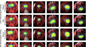
Artificial intelligence assisted operative anatomy recognition in endoscopic pituitary surgery
- Danyal Z. Khan
- Alexandra Valetopoulou
- Hani J. Marcus
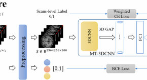
Improved patient identification by incorporating symptom severity in deep learning using neuroanatomic images in first episode schizophrenia
- Wenjing Zhang
- Lituan Wang
- Qiyong Gong
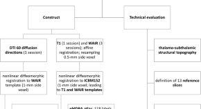
An MRI Deep Brain Adult Template With An Advanced Atlas-Based Tool For Diffusion Tensor Imaging Analysis
- Jean-Jacques Lemaire
- Denys Fontaine
News and Comment

Stress can disrupt memory and lead to needless anxiety — here’s how
In mice, stress altered the way that the brain formed memories, resulting in an unnecessary fear response.
- Smriti Mallapaty
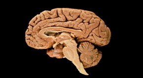
How human brains got so big: our cells learned to handle the stress that comes with size
Understanding how human neurons cope with the energy demands of a large, active brain could open up new avenues for treating neurological disorders.
- Miryam Naddaf

Should Alzheimer’s be diagnosed with a blood test? Proposal sparks controversy
Proponents say that early diagnosis is beneficial. But opponents fear distress for people who might never develop symptoms.

Why do wet dogs shake themselves dry? Neuroscience has an answer
Deciphering how mammals respond to sensations through their fur could inspire further research on skin sensitivity.

What’s so special about the human brain? A graphical guide
Torrents of data from cell atlases, brain organoids and other methods are finally delivering answers to an age-old question.
- Kerri Smith
- Nik Spencer

The brain summons deep sleep for healing from life-threatening injury
A heart attack unleashes immune cells that stimulate sleep neurons, leading to restorative slumber.
- Mariana Lenharo

Quick links
- Explore articles by subject
- Guide to authors
- Editorial policies

Transforming the understanding and treatment of mental illnesses.
Información en español
- Science News
- Meetings and Events
- Social Media
- Press Resources
- Email Updates
- Innovation Speaker Series
Scientists Unveil Detailed Cell Maps of the Human Brain and the Nonhuman Primate Brain
Incredibly detailed cell maps help pave the way for new generation of treatments
October 12, 2023 • Press Release
A group of international scientists have mapped the genetic, cellular, and structural makeup of the human brain and the nonhuman primate brain. This understanding of brain structure, achieved by funding through the National Institutes of Health’s Brain Research Through Advancing Innovative Neurotechnologies ® Initiative, or The BRAIN Initiative® , allows for a deeper knowledge of the cellular basis of brain function and dysfunction, helping pave the way for a new generation of precision therapeutics for people with mental disorders and other disorders of the brain. The findings appear in a compendium of 24 papers across Science , Science Advances , and Science Translational Medicine .
“Mapping the brain’s cellular landscape is a critical step toward understanding how this vital organ works in health and disease,” said Joshua A. Gordon, M.D., Ph.D. , director of the National Institute of Mental Health. “These new detailed cell atlases of the human brain and the nonhuman primate brain offer a foundation for designing new therapies that can target the specific brain cells and circuits involved in brain disorders.”
The 24 papers in this latest BRAIN Initiative Cell Census Network (BICCN) collection detail the exceptionally complex diversity of cells in the human brain and the nonhuman primate brain. The studies identify similarities and differences in how cells are organized and how genes are regulated in the human brain and the nonhuman primate brain. For example:
- Three papers in the collection present the first atlas of cells in the adult human brain, mapping the transcriptional and epigenomic landscape of the brain. The transcriptome is the complete set of gene readouts in a cell, which contains instructions for making proteins and other cellular products. The epigenome refers to chemical modifications to a cell’s DNA and chromosomes that alter the way the cell’s genetic information is expressed.
- In another paper, a comparison of the cellular and molecular properties of the human brain and several nonhuman primate brains (chimpanzee, gorilla, macaque, and marmoset brains) revealed clear similarities in the types, proportions, and spatial organization of cells in the cerebral cortex of humans and nonhuman primates. Examination of the genetic expression of cortical cells across species suggests that relatively small changes in gene expression in the human lineage led to changes in neuronal wiring and synaptic function that likely allowed for greater brain plasticity in humans, supporting the human brain’s ability to adapt, learn, and change.
- A study exploring how cells vary in different brain regions in marmosets found a link between the properties of cells in the adult brain and the properties of those cells during development. The link suggests that developmental programming is embedded in cells when they are formed and maintained into adulthood and that some observable cellular properties in an adult may have their origins very early in life. This finding could lead to new insights into brain development and function across the lifespan.
- An exploration of the anatomy and physiology of neurons in the outermost layer of the neocortex—part of the brain involved in higher-order functions such as cognition, motor commands, and language—revealed differences in the human brain and the mouse brain that suggest this region may be an evolutionary hotspot, with changes in humans reflecting the higher demands of regulating humans’ more complex brain circuits.
The core aim of the BICCN, a groundbreaking effort to understand the brain’s cellular makeup, is to develop a comprehensive inventory of the cells in the brain—where they are, how they develop, how they work together, and how they regulate their activity—to better understand how brain disorders develop, progress, and are best treated.
“This suite of studies represents a landmark achievement in illuminating the complexity of the human brain at the cellular level,” said John Ngai, Ph.D. , director of the NIH BRAIN Initiative. “The scientific collaborations forged through BICCN are propelling the field forward at an exponential pace; the progress—and possibilities—have been simply breathtaking.”
The census of brain cell types in the human brain and the nonhuman primate brain presented in this paper collection serves as a key step toward developing the brain treatments of the future. The findings also set the stage for the BRAIN Initiative Cell Atlas Network , a transformative project that, together with two other large-scale projects—the BRAIN Initiative Connectivity Across Scales and the Armamentarium for Precision Brain Cell Access —aim to revolutionize neuroscience research by illuminating foundational principles governing the circuit basis of behavior and informing new approaches to treating human brain disorders.
Maroso, M. (2023). A quest into the human brain. Science . http://www.science.org/doi/10.1126/science.adl0913
Projects funded through the NIH BRAIN Initiative Cell Census Network
About the National Institute of Mental Health (NIMH): The mission of the NIMH is to transform the understanding and treatment of mental illnesses through basic and clinical research, paving the way for prevention, recovery and cure. For more information, visit the NIMH website .
The NIH BRAIN Initiative is managed by 10 Institutes and Centers whose missions and current research portfolios complement the goals of The BRAIN Initiative®: National Center for Complementary and Integrative Health, National Eye Institute, National Institute on Aging, National Institute on Alcohol Abuse and Alcoholism, National Institute of Biomedical Imaging and Bioengineering, Eunice Kennedy Shriver National Institute of Child Health and Human Development, National Institute on Drug Abuse, National Institute on Deafness and other Communication Disorders, National Institute of Mental Health, and National Institute of Neurological Disorders and Stroke.
About the National Institutes of Health (NIH) : NIH, the nation's medical research agency, includes 27 Institutes and Centers and is a component of the U.S. Department of Health and Human Services. NIH is the primary federal agency conducting and supporting basic, clinical, and translational medical research, and is investigating the causes, treatments, and cures for both common and rare diseases. For more information about NIH and its programs, visit the NIH website .
NIH…Turning Discovery Into Health ®
- U.S. Department of Health & Human Services

- Virtual Tour
- Staff Directory
- En Español
You are here
Nih research matters.
October 31, 2023
Scientists build largest maps to date of cells in human brain
At a glance.
- International research teams created highly detailed cellular maps of adult and developing human brains, along with the brains of other animals.
- These comprehensive cell atlases could help lead to new insights for improving treatments for a host of mental conditions and brain disorders.

The human brain is made up of about 86 billion nerve cells, along with many other types of cells. They interact and link together in unique ways, creating distinct brain regions with specific functions. Uncovering the complex makeup and interactions of these many cells could lead to a new understanding of how the brain functions in health and disease, and new tools to study the complex activities and functions of these cells.
To better understand the identities and roles of brain cells, NIH’s Brain Research Through Advancing Innovative Neurotechnologies® (BRAIN) Initiative launched an international network of collaborating researchers called the BRAIN Initiative Cell Census Network. Its aim is to create a comprehensive inventory of all the cells in the human, nonhuman primate, and mouse brains, including cell locations, interconnections, and activities. The study of brain cells across species can pinpoint features that are uniquely human and give insights into which animals to study for different scientific questions. The latest findings were reported in a series of more than 20 papers published in Science, Science Advances, and Science Translational Medicine on October 13, 2023.
One paper examined three human brains to find over 3,000 types of brain cells—more than previously known. The team identified specific types of cells in distinct clusters in different brain regions. These findings could help shed light on conditions that are known to affect specific brain areas, such as cancer or neurodegenerative diseases.
The researchers have created the most detailed cell atlas yet of the adult human brain. The atlas reveals information about each cell’s gene activity and epigenome—the changes to a cell’s DNA and chromosomes that alter genetic activity. The findings also show that, besides variation among brain regions, there is variation between individuals. More people will need to be studied to fully understand the patterns of healthy and diseased brains.
Another paper compared the cellular and molecular properties of the brains of humans and several nonhuman primates: the chimpanzee, gorilla, macaque, and marmoset. The scientists found that a few hundred genes had activity patterns in nerve cells that were unique to humans. These changes might help to explain humans’ remarkable ability to adapt, learn, and change.
The other papers covered various aspects of the brain. One, for example, explored the role that inflammation might play during early brain development. Severe inflammation in childhood has been linked to developmental disorders like autism and schizophrenia. Researchers analyzed gene activity in the brains of children who died when they were 1 to 5 years old. They compared the brains of children who died with inflammatory conditions, such as asthma or infections, to those who died from accidents. The scientists focused on the brain’s cerebellum, which controls muscle movement and cognitive functions like language and social skills. They found evidence that inflammation can block development of specific types of nerve cells in the cerebellum. The finding could lead to better understanding and treatment of developmental disorders that are linked to inflammation.
“This suite of studies represents a landmark achievement in illuminating the complexity of the human brain at the cellular level,” says Dr. John Ngai, director of the NIH BRAIN Initiative.
“These new detailed cell atlases of the human brain and the nonhuman primate brain offer a foundation for designing new therapies that can target the specific brain cells and circuits involved in brain disorders,” adds Dr. Joshua A. Gordon, director of NIH’s National Institute of Mental Health.
Related Links
- Cell Atlases Give Detailed Views of Human Organs
- Mapping the Mammalian Motor Cortex
- Building an Atlas of Brain Function in Mice
- An Expanded Map of the Human Brain
- An Atlas of the Developing Human Brain
- Get To Know Your Brain
- Brain Basics: Know Your Brain
- NIH’s BRAIN Initiative
References: A quest into the human brain. Maroso M . Science . 2023 Oct 13; 382(6667): 166-167. doi: 10.1126/science.adl0913. Epub 2023 Oct 12. PMID: 37824675. Transcriptomic diversity of cell types across the adult human brain. Siletti K, Hodge R, Mossi Albiach A, Lee KW, Ding SL, Hu L, Lönnerberg P, Bakken T, Casper T, Clark M, Dee N, Gloe J, Hirschstein D, Shapovalova NV, Keene CD, Nyhus J, Tung H, Yanny AM, Arenas E, Lein ES, Linnarsson S. Science. 2023 Oct 13;382(6667):eadd7046. doi: 10.1126/science.add7046. Epub 2023 Oct 13. PMID: 37824663. Comparative transcriptomics reveals human-specific cortical features. Jorstad NL, Song JHT, Exposito-Alonso D, Suresh H, Castro-Pacheco N, Krienen FM, Yanny AM, Close J, Gelfand E, Long B, Seeman SC, Travaglini KJ, Basu S, Beaudin M, Bertagnolli D, Crow M, Ding SL, Eggermont J, Glandon A, Goldy J, Kiick K, Kroes T, McMillen D, Pham T, Rimorin C, Siletti K, Somasundaram S, Tieu M, Torkelson A, Feng G, Hopkins WD, Höllt T, Keene CD, Linnarsson S, McCarroll SA, Lelieveldt BP, Sherwood CC, Smith K, Walsh CA, Dobin A, Gillis J, Lein ES, Hodge RD, Bakken TE. Science. 2023 Oct 13;382(6667):eade9516. doi: 10.1126/science.ade9516. Epub 2023 Oct 13. PMID: 37824638. A single-cell genomic atlas for maturation of the human cerebellum during early childhood. Ament SA, Cortes-Gutierrez M, Herb BR, Mocci E, Colantuoni C, McCarthy MM. Sci Transl Med . 2023 Oct 12:eade1283. doi: 10.1126/scitranslmed.ade1283. Online ahead of print. PMID: 37824600.
Funding: NIH’s BRAIN Initiative Cell Census Network (BICCN).
Connect with Us
- More Social Media from NIH
Suggestions or feedback?
MIT News | Massachusetts Institute of Technology
- Machine learning
- Sustainability
- Black holes
- Classes and programs
Departments
- Aeronautics and Astronautics
- Brain and Cognitive Sciences
- Architecture
- Political Science
- Mechanical Engineering
Centers, Labs, & Programs
- Abdul Latif Jameel Poverty Action Lab (J-PAL)
- Picower Institute for Learning and Memory
- Lincoln Laboratory
- School of Architecture + Planning
- School of Engineering
- School of Humanities, Arts, and Social Sciences
- Sloan School of Management
- School of Science
- MIT Schwarzman College of Computing
Study reveals a universal pattern of brain wave frequencies
Press contact :, media download.
*Terms of Use:
Images for download on the MIT News office website are made available to non-commercial entities, press and the general public under a Creative Commons Attribution Non-Commercial No Derivatives license . You may not alter the images provided, other than to crop them to size. A credit line must be used when reproducing images; if one is not provided below, credit the images to "MIT."
Previous image Next image
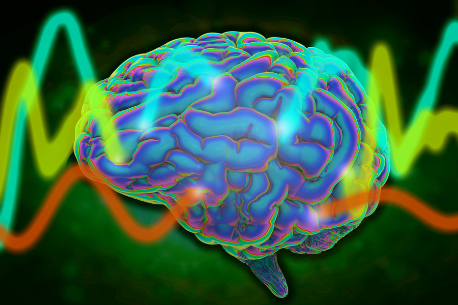
Throughout the brain’s cortex, neurons are arranged in six distinctive layers, which can be readily seen with a microscope. A team of MIT and Vanderbilt University neuroscientists has now found that these layers also show distinct patterns of electrical activity, which are consistent over many brain regions and across several animal species, including humans.
The researchers found that in the topmost layers, neuron activity is dominated by rapid oscillations known as gamma waves. In the deeper layers, slower oscillations called alpha and beta waves predominate. The universality of these patterns suggests that these oscillations are likely playing an important role across the brain, the researchers say.

“When you see something that consistent and ubiquitous across cortex, it’s playing a very fundamental role in what the cortex does,” says Earl Miller, the Picower Professor of Neuroscience, a member of MIT’s Picower Institute for Learning and Memory, and one of the senior authors of the new study.
Imbalances in how these oscillations interact with each other may be involved in brain disorders such as attention deficit hyperactivity disorder, the researchers say.
“Overly synchronous neural activity is known to play a role in epilepsy, and now we suspect that different pathologies of synchrony may contribute to many brain disorders, including disorders of perception, attention, memory, and motor control. In an orchestra, one instrument played out of synchrony with the rest can disrupt the coherence of the entire piece of music,” says Robert Desimone, director of MIT’s McGovern Institute for Brain Research and one of the senior authors of the study.
André Bastos, an assistant professor of psychology at Vanderbilt University, is also a senior author of the open-access paper, which appears today in Nature Neuroscience . The lead authors of the paper are MIT research scientist Diego Mendoza-Halliday and MIT postdoc Alex Major.
Layers of activity
The human brain contains billions of neurons, each of which has its own electrical firing patterns. Together, groups of neurons with similar patterns generate oscillations of electrical activity, or brain waves, which can have different frequencies. Miller’s lab has previously shown that high-frequency gamma rhythms are associated with encoding and retrieving sensory information, while low-frequency beta rhythms act as a control mechanism that determines which information is read out from working memory.
His lab has also found that in certain parts of the prefrontal cortex, different brain layers show distinctive patterns of oscillation: faster oscillation at the surface and slower oscillation in the deep layers. One study , led by Bastos when he was a postdoc in Miller’s lab, showed that as animals performed working memory tasks, lower-frequency rhythms generated in deeper layers regulated the higher-frequency gamma rhythms generated in the superficial layers.
In addition to working memory, the brain’s cortex also is the seat of thought, planning, and high-level processing of emotion and sensory information. Throughout the regions involved in these functions, neurons are arranged in six layers, and each layer has its own distinctive combination of cell types and connections with other brain areas.
“The cortex is organized anatomically into six layers, no matter whether you look at mice or humans or any mammalian species, and this pattern is present in all cortical areas within each species,” Mendoza-Halliday says. “Unfortunately, a lot of studies of brain activity have been ignoring those layers because when you record the activity of neurons, it's been difficult to understand where they are in the context of those layers.”
In the new paper, the researchers wanted to explore whether the layered oscillation pattern they had seen in the prefrontal cortex is more widespread, occurring across different parts of the cortex and across species.
Using a combination of data acquired in Miller’s lab, Desimone’s lab, and labs from collaborators at Vanderbilt, the Netherlands Institute for Neuroscience, and the University of Western Ontario, the researchers were able to analyze 14 different areas of the cortex, from four mammalian species. This data included recordings of electrical activity from three human patients who had electrodes inserted in the brain as part of a surgical procedure they were undergoing.
Recording from individual cortical layers has been difficult in the past, because each layer is less than a millimeter thick, so it’s hard to know which layer an electrode is recording from. For this study, electrical activity was recorded using special electrodes that record from all of the layers at once, then feed the data into a new computational algorithm the authors designed, termed FLIP (frequency-based layer identification procedure). This algorithm can determine which layer each signal came from.
“More recent technology allows recording of all layers of cortex simultaneously. This paints a broader perspective of microcircuitry and allowed us to observe this layered pattern,” Major says. “This work is exciting because it is both informative of a fundamental microcircuit pattern and provides a robust new technique for studying the brain. It doesn’t matter if the brain is performing a task or at rest and can be observed in as little as five to 10 seconds.”
Across all species, in each region studied, the researchers found the same layered activity pattern.
“We did a mass analysis of all the data to see if we could find the same pattern in all areas of the cortex, and voilà, it was everywhere. That was a real indication that what had previously been seen in a couple of areas was representing a fundamental mechanism across the cortex,” Mendoza-Halliday says.
Maintaining balance
The findings support a model that Miller’s lab has previously put forth, which proposes that the brain’s spatial organization helps it to incorporate new information, which carried by high-frequency oscillations, into existing memories and brain processes, which are maintained by low-frequency oscillations. As information passes from layer to layer, input can be incorporated as needed to help the brain perform particular tasks such as baking a new cookie recipe or remembering a phone number.
“The consequence of a laminar separation of these frequencies, as we observed, may be to allow superficial layers to represent external sensory information with faster frequencies, and for deep layers to represent internal cognitive states with slower frequencies,” Bastos says. “The high-level implication is that the cortex has multiple mechanisms involving both anatomy and oscillations to separate ‘external’ from ‘internal’ information.”
Under this theory, imbalances between high- and low-frequency oscillations can lead to either attention deficits such as ADHD, when the higher frequencies dominate and too much sensory information gets in, or delusional disorders such as schizophrenia, when the low frequency oscillations are too strong and not enough sensory information gets in.
“The proper balance between the top-down control signals and the bottom-up sensory signals is important for everything the cortex does,” Miller says. “When the balance goes awry, you get a wide variety of neuropsychiatric disorders.”
The researchers are now exploring whether measuring these oscillations could help to diagnose these types of disorders. They are also investigating whether rebalancing the oscillations could alter behavior — an approach that could one day be used to treat attention deficits or other neurological disorders, the researchers say.
The researchers also hope to work with other labs to characterize the layered oscillation patterns in more detail across different brain regions.
“Our hope is that with enough of that standardized reporting, we will start to see common patterns of activity across different areas or functions that might reveal a common mechanism for computation that can be used for motor outputs, for vision, for memory and attention, et cetera,” Mendoza-Halliday says.
The research was funded by the U.S. Office of Naval Research, the U.S. National Institutes of Health, the U.S. National Eye Institute, the U.S. National Institute of Mental Health, the Picower Institute, a Simons Center for the Social Brain Postdoctoral Fellowship, and a Canadian Institutes of Health Postdoctoral Fellowship.
Share this news article on:
Related links.
- Robert Desimone
- Earl Miller
- McGovern Institute for Brain Research
- Department of Brain and Cognitive Sciences
Related Topics
- Brain and cognitive sciences
- Neuroscience
- McGovern Institute
- Picower Institute
- National Institutes of Health (NIH)
Related Articles

“Spatial computing” enables flexible working memory
Controlling attention with brain waves
New study reveals how brain waves control working memory

How the brain pays attention
Previous item Next item
More MIT News

Building an understanding of how drivers interact with emerging vehicle technologies
Read full story →

Consortium led by MIT, Harvard University, and Mass General Brigham spurs development of 408 MW of renewable energy

A vision for U.S. science success

Catherine Wolfram: High-energy scholar

MIT researchers develop an efficient way to train more reliable AI agents

Advancing urban tree monitoring with AI-powered digital twins
- More news on MIT News homepage →
Massachusetts Institute of Technology 77 Massachusetts Avenue, Cambridge, MA, USA
- Map (opens in new window)
- Events (opens in new window)
- People (opens in new window)
- Careers (opens in new window)
- Accessibility
- Social Media Hub
- MIT on Facebook
- MIT on YouTube
- MIT on Instagram

IMAGES
COMMENTS
Published in Science, the study is the latest development in a nearly 10-year collaboration with scientists at Google Research, combining Lichtman’s electron microscopy imaging with AI algorithms to color-code and reconstruct the extremely complex wiring of mammal brains.
Researchers generated a high-resolution map of all the cells and connections in a single cubic millimeter of the human brain. The results reveal previously unseen details of brain structure and provide a resource for further studies. A single neuron is shown with 5,600 of the nerve fibers (blue) that connect to it.
Understanding how human neurons cope with the energy demands of a large, active brain could open up new avenues for treating neurological disorders. Should Alzheimer’s be diagnosed with a blood...
A group of international scientists have mapped the genetic, cellular, and structural makeup of the human brain and the nonhuman primate brain, allowing for a deeper knowledge of the cellular basis of brain function and dysfunction, helping pave the way for a new generation of precision therapeutics for people with mental disorders and other ...
International research teams created highly detailed cellular maps of adult and developing human brains, along with the brains of other animals. These comprehensive cell atlases could help lead to new insights for improving treatments for a host of mental conditions and brain disorders.
The six anatomical layers of the mammalian brain cortex show distinct patterns of electrical activity which are consistent throughout the entire cortex and across several animal species, including humans, an MIT study has found.